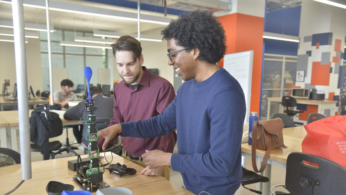
Design, Collaborate, MAKE
With locations on Main Campus and Downtown Campus, our Makerspaces include resources for all students to use to work on their projects while sharing ideas, equipment and knowledge.
30
Degree programs
4200
Students
143
Faculty
Latest News
Read All News Stories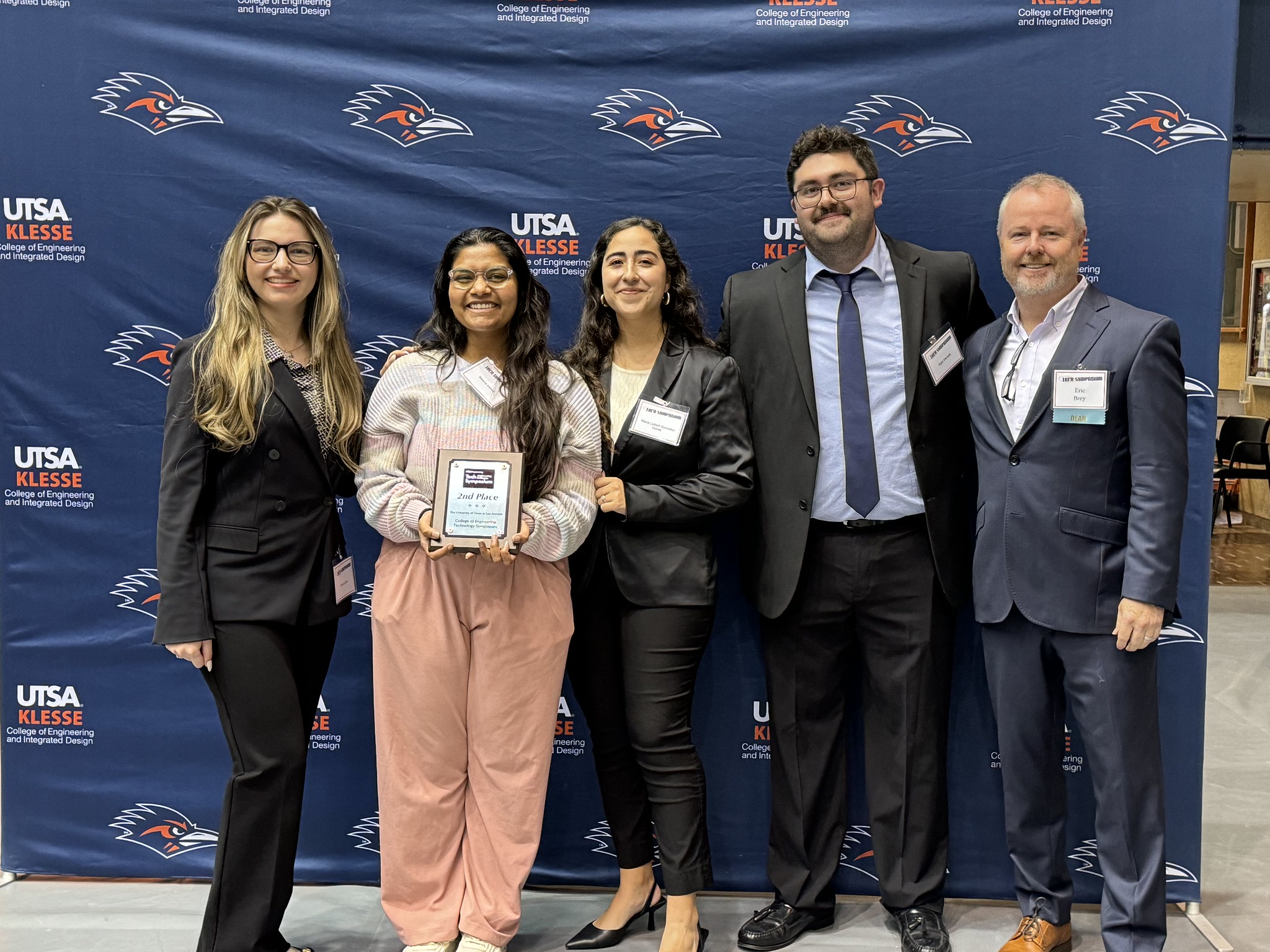
April 26, 2024
Device for detecting retinal disease takes top prize in UTSA’s spring Tech SymposiumPublished by Katrina Kehoe
April 18, 2024
Professors Use Machine Learning for Model to Predict Demolition Orders and Assess Code Enforcement BiasPublished by Sean M. Wood
Klesse College Events
View All EventsMay
15
2024
Order of the Engineer CeremonyGraduating Spring with a degree in Engineering at UTSA? Register to attend the Order of the Engineer Ceremony. Friends and family welcome to attend!
May
20 - 22
2024
American Society of Civil Engineers - Utility Engineering and Surveying Institute Texas Section 5th Annual ConferenceUtilities in Texas: Past, Present, & Future
Jul
26 - 27
2024
4th Annual Texas Utility Engineering and Surveying Conference hosted by CUIRE/UTAIncludes Keynote Presentation and Three Concurrent Tracks, Exhibits, Lunch and Refreshments.